Dendrites are branched filaments in nerve cells (neurons). The word dendrite dervives from the Greek word for tree which describes their branching tree-like structure. Here is a diagram of a typical neuron showing the position of the dendrites:
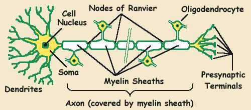
A typical neuron in the central nervous system (CNS) has many thousand dendrites. It is the dendrites that receive the bulk of nervous signals from other neurons via connections called synapses. Here is a diagram of some synapses:
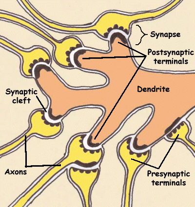
Dendrites provide the postsynaptic terminal component of a synapse. The nerve transmissions are received from the axons of other neurons via small signalling molecules called neurotransmitters.
Dendrites differ from axons in a number of ways:
Dendrites carry nerve impulses to the neuronal cell body (the soma) whereas the axons carry it away.
A neuron has very many dendrites but typically only one axon (although it can have more than one).
Dendrites are much thicker than synapses.
Some axons are sheathed in an insulating fatty protein called myelin. Dendites are not sheathed in myelin.
Dendrites are short whereas axons are long - sometimes as long as a metre or more.
Dendrite links:
Structure of the Neuron - Dendrites
Source details
Author: Paul Jones
Archive capture: . This is not a medical review date.